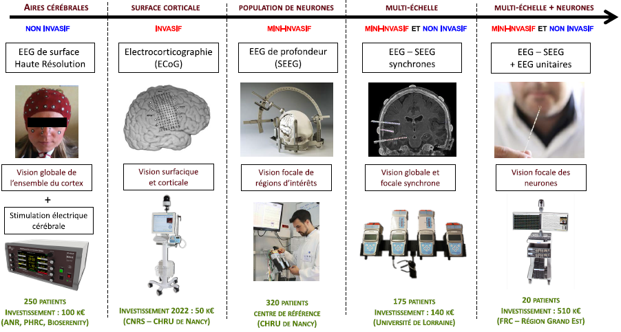
This platform provides access to in vivo recording in humans (and more particularly in epileptic patients) of brain electrical activity at different scales. The dynamics of brain processes currently require investigation methods whose temporal resolution must be of the order of milliseconds. Electroencephalography (EEG) is one of the only methods that meets this criterion. The study of brain function can be carried out at different spatial scales: networks of neurons that use surface EEG, populations of neurons that require intracerebral EEG (also called Stereo-EEG) or unit neurons that require unit EEG (also called Single-unit recording). At the CRAN, in collaboration with the CHRU of Nancy, we benefit on the same site from all these EEG methods. The other original aspect is based on the development in Nancy since 2016 of unit EEG. For this type of unit EEG recording in vivo in humans, we will take advantage of intra-cerebral EEG recordings using macro-contacts probes (Stereo-ElectroEncephaloGraphy: SEEG) prescribed and performed in clinical routine in patients with partial epilepsy refractory to drugs . These explorations will be part of a pre-surgical assessment and will be carried out in the Neurology Department of the University Hospital of Nancy.
The first scientific objective is to evaluate the specific face detection capabilities of individual neurons compared to their object detection capabilities in the medial temporal lobe and in the ventral temporal cortex. The second objective is to understand the functioning of physiological processes at different scales. The links between these signals measured at different spatial scales are far from being understood, especially at the level of the relationships between microelectrode recordings and macroelectrodes.
From a methodological point of view, unit and clinical EEG recordings are coupled with periodic visual stimulations that generate electrical evoked potentials in cortical areas selectively activated by defined cognitive protocols.
The 256-channel acquisition system is coupled with video allowing simultaneous measurement by micro and macro-contacts of signals generated by a single neuron and by a population of neurons. All measurements obtained on the 256 channels are synchronized to study the relationships between the signals obtained at different anatomical scales (neurons versus populations of neurons). The probes to be implanted by neurosurgery contain micro-contacts with adjustable exploration depth (wires with a diameter of 25 μm) and conventional macro-contacts (with a diameter of 0.8 mm) such as those used in clinical routine. The amplifiers of the neuronal signals have remarkable characteristics such as amplifying signals of a few nanovolts with a common mode rejection rate greater than 90 dB at 50 Hz. If the sampling frequencies of the SEEG acquisition systems can be limited to 4 KHz, the sampling frequency for unit activity recording can go up to 30 KHz.
A research platform is associated with this video-EEG recording device in order to be able to:
1. perform cognitive periodic visual stimulations during EEG recordings
2. process/interpret the signals collected in a signal processing system for clinical and research purposes
3. share the signals between the CHU of Nancy and our laboratory via a secure internet network.