— Development of 2D or 3D co-culture models to evaluate the impact of therapy on a complex model of tumor cells and cells of the microenvironment + Evaluation of the recruitment of microenvironment cells according to treatment conditions. (Ligue Sophie Pinel project, Multi-scale modeling and characterization of post-radiotherapy recurrences of glioblastomas, 2021– ).
— Characterize the infiltration and polarization of macrophages post-treatment on 3D culture models (Euronanomed III RXnanoBRAIN project, Muriel Barbery-Heyob, 2021–);
— Fusion of multimodal information and numerical modeling of the effects of modulation of aromatase expression on glioma development (INS2I Emergence project, InfoGlio, Hélène Dumond, 2020–);
— Characterize the effect of GPER agonist on cell morphology and functions (proliferation, migration. . .) in a 2D culture model and invasion in an organotypic culture model (3D, 2019–);
— Development of numerical models of the biological response of a glioblastoma to radiation therapy in the presence of nanoparticles, studying not only the effects on tumor growth, but also on the invasion of tumor cells (E.-H. Djermoune, M. Thomassin, 2019–);
— Evaluation of new peptide-based biomarkers c(RGDfK) targeting integrins in a brain tumor model (ANR Modality project Cédric Boura, 2018–2021);
— Evaluate the impact of hybrid gold-gadolinium nanoparticles on the migratory and invasive capacities of irradiated or non-irradiated glial tumor cells: analyses in endpoint or dynamic (e-NanoRX project, Sophie Pinel, 2017–2021).
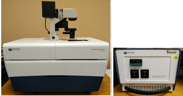
The ImageXpress Micro Confocal High-content is an automated and ultra-fast imaging system that allows rapid widefield and confocal imaging of living or fixed samples at a fixed time (end-point) or at regular intervals (time-lapse). Equipped with different objectives (4X, 10X, 20X, 40X and 60X), this equipment allows acquisition not only in brightfield mode but also in fluorescence mode (FITC, Cy5, Cy3, DAPI and Texas red; with independent excitation of the emission). The MetaXPress analysis software includes different basic modules for quick and guided analyses (application module). It also has all the tools necessary to allow more advanced and custom analysis (custom module).