The BioSiS department of the CRAN conducts research in oncology, particularly on the interactions between radiation and biological tissues. This axis is highly interdisciplinary, bringing together biologists, signal processors, radiophysicists, and clinicians whose areas of expertise are very complementary. In this axis, the objectives are:
— to optimize and evaluate, on preclinical models, alternative and innovative therapeutic strategies, combining nanoparticles and radiation (ionizing or non-ionizing), to treat adult and pediatric brain tumors;
— to model and control in real time the effects of radiation therapies on tumor growth or recurrence.
The recognition of the expertise of this project in the fields of adult and pediatric brain tumors, radiotherapy, and nanoparticles is reflected in the active involvement in various learned societies and the obtaining of national and international projects.
The laboratory has a preclinical irradiator equipped with planar imaging equipment. This platform is part of a voluntary mutualization approach. Pricing rules have been defined and published in the CNRS B.O. The governance of the platform has been established and disseminated, stipulating in particular that access to the devices is conditional on the prior submission of a project sheet. Each project leads to the creation of a specific account allowing precise monitoring of the use of the equipment. The pricing applied since August 1, 2020 has been published in the CNRS B.O. (B.O. of December 2020).
Recently, several external teams have been using it:
— Cell Therapy Unit (UTCT), CHU Nancy: 4 projects open since August;
— IMoPA: 1 project submitted in November 2020;
— IADI – radiochemistry: 1 project submitted.
Billing (semi-annual) is based on the hourly statements of each user account, in accordance with the platform’s regulations.
Acquired in 2015, the X-RAD320 irradiator (Precision X-ray inc.), which delivers X-ray photons with energies between 11 kV and 320 keV, allows the irradiation of monolayer or three-dimensional cell cultures and the focused irradiation of rodents (mice, rats). A motorized collimator allows irradiation of fields from 1×1 cm2 to 20×20 cm2. The generator is a General Electric Titan Isovolt E 320.
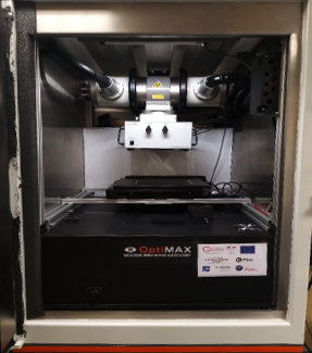
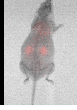
Since the end of 2018, a multimodal imaging module (bioluminescence / scintigraphy / X) allows the implementation of image-guided radiotherapy protocols or analysis and monitoring of specific tumor models. This module (OptiMAX, Precision X-ray inc., North Branford, CT) consists of a CCD camera cooled to -80°C. The sensor has 1024×1024 elements of 13 μm per side. The conversion of X-ray photons into light photons is ensured by a retractable phosphor film. Thus, the same camera can acquire bioluminescence and radiographic images, or even scintigraphic images if the radioelement used emits photons with energy less than 150 keV. For higher energies, the phosphor film is ineffective.
A computer engineer was recruited through the INS2I 2020 call for projects for the development of tomographic reconstruction algorithms for the short-term evolution of the equipment.
In 2020, a confidentiality agreement was signed between the University of Lorraine (represented by the CRAN) and Precision X-ray inc. (North Brandford, CT) around the developments of X / bioluminescence preclinical tomography.
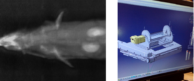
As part of a local call for tenders at the laboratory, a tomographic insert is being produced by the research support service of the unit. This insert allows the acquisition of morphological (X) and functional (bioluminescence / scintigraphy) three-dimensional images. The prototype is equipped with a motorized translation stage on 2 axes to position the area to be irradiated based on the acquired morphological and/or functional images; thus allowing true image-guided radiotherapy (IGRT).