Acquisition and formatting of data; development of segmentation, identification, classification, knowledge modeling, and automatic contour generation methods, hyperspectral image processing (unmixing, deconvolution, fusion)
The multimodal imaging platform (PIM) project brings together in one place a unique set of industrial imaging instruments. It aims to allow the Signal Image Vision community of the GDR ISIS and the production system control community of the GDR MACS to access this set of instruments by taking charge of the production of experimental data acquired under controlled conditions according to a protocol designed and defined by potential users. This data must allow testing and validating image processing and computer vision models and algorithms. As examples, we can mention:
— hyperspectral imaging (unmixing, classification, super-resolution, deconvolution);
— 3D reconstruction from RGB-D camera;
— 3D reconstruction in X-ray tomography;
— 3D reconstruction of point clouds obtained by terrestrial LIDAR;
— multimodal data fusion;
— the contribution of multimodality to texture characterization;
— the contribution of multimodality to non-destructive testing.
A website details the technical characteristics of the equipment, the management of acquisition or processing requests on site or remotely, and the governance arrangements. This platform is integrated into the TRACILOGIS network (Traceability, Identification, intelligent control for the wood supply chain) by providing non-destructive testing tools to identify and evaluate product quality, and help decision-making in flow control.
The aim is to be able to analyze how these systems behave under conditions close to those of online material control systems. One of the objectives of the PIM project was to establish a database that will be made available to the GDR ISIS community in connection with Yannick Berthoumieu. Particular focus will be given to the construction of hyperspectral image libraries for 3 specific problems.
Modeling nonlinear phenomena in hyperspectral imaging: a first aspect concerns multiple scattering induced by the geometry of the imaged scene. The aim is to design a set of test samples made with materials with known optical properties and arranged in a geometry allowing control of multiple reflections. For each object, an RGB-D image will also be provided. It will provide access to the topography of the imaged object. A second aspect concerns the study of multiple scattering phenomena within intimate mixtures. The aim is to design and image samples including intimate mixtures (sands), in controlled proportions, of a small number of chemical compounds.
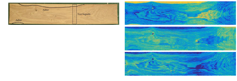
Modeling and characterizing textures of hyperspectral images of wood samples: the objective is to build a library of images of different types of wood, which is an interesting material for texture analysis. Particular focus will be given to the characterization of textures of heartwood (noble wood) and sapwood (porous wood) on different types of wood. For each object, an RGB-D image will also be provided. It will provide a color (visible) image with high spatial resolution.
Sequential processing of hyperspectral images (pushbroom imager): the aim is to acquire data under various experimental conditions (type and size of materials, integration time, conveyor speed, pixel binning) to evaluate their effects (blur, noise) on the observed images and to propose benchmarks to evaluate the performance of processing algorithms.
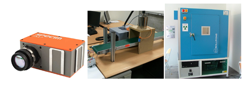
The platform includes four main pieces of equipment:
Hyperspectral linear camera in the near infrared (NIR). The hyperspectral camera acquires a line of 640 pixels. For each of these pixels, light intensity is measured at 224 wavelengths, from 935 to 1702 nm (near infrared). An automated system allows the sample to be moved to scan it. The instrument thus provides a “data cube,” the spectral axis being added to the usual spatial axes.
NIR-FT spectrometer (near infrared Fourier transform) Bruker. the Brucker Matrix F spectrometer is a NIR-FT (near infrared Fourier transform) spectrometer with emission option for contactless measurements. The contactless measuring head has two tungsten sources that illuminate the sample. The scattered light is collected and conducted to the spectrometer by an optical fiber.
Image acquisition platform (RGB color, monochrome, Scatter, and 3D – obtained by laser profilometry) by the SICK Ranger linear camera. The SICK ColorRanger camera is a high-speed linear camera that allows acquisition:
— of a color image (13000 RGB lines per second);
— of an intensity image in grayscale;
— of a laser profile scatter image;
— of a 3D profilometry image (35000 profiles per second).
The test bench is equipped with a belt conveyor that allows objects to pass under the camera, and various light sources: white LED strip (30W) for color and monochrome image acquisition, lasers (25 mW, 656 nm) for scatter and 3D image acquisition.
X-ray tomograph. The tomograph allows the tomographic acquisition of low-density samples (wood, materials based on natural fibers). The samples have a thickness ranging from a few millimeters to about fifteen centimeters along the cutting axis, and a height of up to about fifty centimeters. The tomograph provides a data cube along the three spatial axes, the volume elements (voxels) having a size ranging from a few microns (for the smallest samples) to about a hundred microns (for the largest).